
What does loss of 3rd Cranial Nerve function cause?

Correlation of pupil abnormalities with clinical findings, history and trends is important. Direct eye injuries can produce CN III abnormalities. Pupillary abnormalities can also be chronic. This can lead to problems with eye movement or eyelid opening where pupil function is spared. The deep blood supply of CN III can become impaired by vascular diseases, hypertension or diabetes. Consequently, a mass that presses on the outside of CN III will usually impact the pupillary function first (for example as a result of an aneurysm or raised ICP). By contrast, the CN III control of eye movement and eyelid opening runs deep in the centre of the nerve.

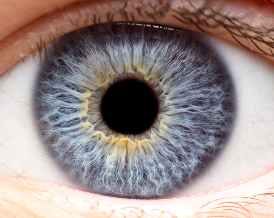
Loss of pupillary reactivity is the most important urgent CN III finding. The pupillary control provided by CN III is located along the periphery of the nerve. A new and sudden finding of pupillary dilation and loss of reactivity suggests supratentorial herniation or expanding volume at the top of the brainstem. What is the significance of monitoring for CN III function?Īcute loss of CN III function is an important sign of a raised intracranial pressure with expanding mass lesion. You can remember this function because the Oculomotor nerve starts with the letter "O" for eye "O"pening. Eyelid Elevation Cranial Nerve III also controls the ability to open the eyelid. CN IV is uncommon, however, difficulty seeing when walking down the stairs is a potential symptom. This portion of the nerve has already crossed. However, if there is compression at the top of the brainstem where CN IV emerges towards the front (ventral) of the brainstem, the dysfunction will be ipsilateral. If the nucleus of the RIGHT CN IV is damaged, downward rotation of the LEFT eye is impaired. It is also the only CN that has contralateral function. At a quick glance, CN IV appears to originate from the top of the brainstem, lateral to CN III. In fact, CN IV is the only CN that originates from the back of the brainstem (dorsal). Thus, both eyes gaze horizontally toward the RIGHT. The stimulus is then carries to the RIGHT CN VI, which activates the LEFT CN III. Turning the head toward the RIGHT stimulates the RIGHT CN VIII (vestibular) involuntarily. Horizontal gaze can be initiated voluntarily (intentionally) or involuntarily. Small or pinpoint pupils may also be present with a lesion in the pons (loss of sympathetic control of the pupil is located in the pons). Inability to move either eye horizontally may indicate injury in the region of the pons or lower brainstem. When the Right CN VI is stimulated, it sends a stimulus up the medial lateral fasciculus to activate the LEFT CN III. The RIGHT EYE rotates horizontally toward the RIGHT temple (Right CN VI) and the LEFT EYE rotates horizontally toward the RIGHT temple (CN III), producing rightward horizontal gaze. CN VI is also ipsilateral (controls the ability to rotate the eye horizontally toward the temple on the same side). Each CN III activates the medial rectus, superior rectus, inferior rectus and inferior oblique muscles to cause orbital rotation on the same side of the CN III nucleus (ipsilateral). The ability to move the eye in all other directions is controlled by CN III. Cranial Nerve IV (Trochlear) controls downward eye movement toward the nose, and Cranial Nerve VI (Abducens) controls horizontal eye movement toward the temple. Eye Movement The 3rd cranial nerve also controls eye muscle movement. The parasympathetic response of the pupil (or "return to normal") is constriction. Pupil Constriction Each one of the two 3rd cranial nerves controls the parasympathetic response of the pupil on the same side (ipsilateral).

They control eye muscles on the same side of the body (ipsilateral). They are Lower Motor Neurons (LMN) (second order neurons). The 3rd cranial nerves are pure motor nerves. The pair of 3rd cranial nerves (oculomotor nerves) are located at the top of the brainstem - one to the right and one to the left.


 0 kommentar(er)
